Dendritic Cells
Dendritic cells (DCs) are professional antigen-presenting cells that act as the link between innate and adaptive immunity. Immature dendritic cells are found in tissues that are in contact with the external environment – such as the skin, and the lining of the nose and lungs – where they sense and sample the environment for self- and non-self antigens. Once activated, they mature and migrate to the lymph nodes, where they present antigen to T cells through major histocompatibility complex (MHC) molecules to start the adaptive immune response.
Formation and structure
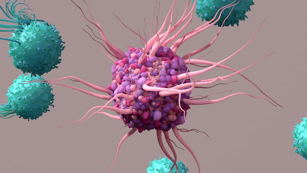
DCs are formed in the bone marrow from hematopoietic stem cells. Immature DCs in peripheral tissue have a round and smooth surface and, as they mature, they display a characteristic tree-like appearance, with large cytoplasmic projections that give the cells their name.
DCs are a heterogeneous group of cells consisting of subsets, including plasmacytoid DCs (pDCs) with a plasma cell-resembling morphology and classical/myeloid DCs (cDCc). cDCs can be further separated into cDC1 and cDC2 types. A 3rd subpopulation formerly seen as CD16+ cDCs1 now has been identified as non-classical monocytes. Each subtype has distinct markers and develops under the control of a specific combination of transcription factors. They differ in location, migratory pathways, and immunological function, however all DCs are capable of antigen uptake, processing and presentation to naïve T cells.
Dendritic cell function – a role in innate and adaptive immunity
pDCs initiate the activation of an adaptive immune response to viruses and play a role in immunoregulation2. Immature cDCs are continuously sampling their environment through a combination of endocytosis, phagocytosis and micropinocytosis. In the absence of inflammatory signals, they are believed to reinforce T cell tolerance. However, when DCs identify pathogens or damage via their pattern recognition receptors (PRRs), they mature and migrate to draining lymph nodes in response to chemokines. As they mature, they undergo a number of phenotypic and functional changes in preparation for activating T lymphocytes. This includes upregulating expression of surface MHC class II and co-stimulatory molecules, as well as pro-inflammatory cytokines. In the lymphoid tissue, they present antigens through MHC class II molecules to CD4+ cells, and cross-present to CD8+ cells through MHC class I molecules, initiating the adaptive immune response.
Emerging research
DCs are complex to study due to their heterogenicity and complex interactions. However, they have an important role in the immune system, and there is great interest in studying them in order to develop therapies targeting autoimmune diseases3 and transplantation intolerance2.
The role of dendritic cells in cancer immunology and immunotherapy is of particular interest, since DCs have been shown to acquire, process, and present tumor-associate antigens in the tumor microenvironment (TME)4. In addition, the TME also contains a network of immunosuppressive factors that can inhibit DC infiltration and subdue their antitumor activity. Targeting these immunosuppressive pathways therapeutically may improve the recruitment, infiltration and effector activity of T cells in the TME.
Tools to study dendritic cells
Cell isolation and culture
DCs can be isolated in small numbers from blood. Alternatively, if larger numbers are required, they can be differentiated from peripheral blood monocytes or generated from human monocytic cell lines.
Functional assays
Assessment of activation of dendritic cells can be used to study the effect of agonists and antagonists on the immune response controlled by dendritic cells, for example in the study of biotherapeutics targeting the innate immune system. Maturation can be measured by looking at upregulation of MHC class I and II, and supernatant samples can be analyzed to study the secretion of cytokines and chemokines.
Cell markers
Most knowledge about the different DC subsets has come from studies in mice, where several lymphoid tissue-resident and migratory DC subsets have been characterized. However, due to significant differences in the cell surface markers expressed by mouse and human DCs, the alignment of phenotypic and functional characteristics between mouse and human DC subsets has been difficult.
There is no single cell marker that is known to be expressed exclusively by DCs, and so a combination of the presence and absence of various proteins can be used to identify them. A combination in expression of CD1c, CD11c, CD16, CD45, CD141, CD370 (CLEC9A), HLA-DR, and lineage exclusion markers CD3, CD14, CD19, CD20, CD56 can be used to differentiate between the main subsets and other types of cells.
| Cell Marker | Alternative Names |
|---|---|
| cDC1 | |
| HLA-DR | |
| CD11c | Integrin alpha-X, ITGAX, SLEB6 |
| CD26 | DPP4, ADABP, ADCP2, DPPIV, TP103 |
| CD141 | Thrombomodulin, THBD, THRM, BDCA-3 |
| CD226 | DNAM-1, PTA1 |
| CD272 | BTLA |
| CD370 | CLEC9A, HEEE9341, UNQ9341, DNGR1 |
| CADM1 | L2, IGSF4, IGSF4A, NECL2, Necl-2, RA175, ST17, SYNCAM, TSLC1, sTSLC-1, sgIGSF, synCAM1, cell adhesion molecule 1 |
| XCR1 | CCGPR5, X-C motif chemokine receptor 1 |
| cDC2 | |
| HLA-DR | |
| CD1c | BDCA-1 |
| CD2 | SRBC, T11 |
| CD11b | Integrin alpha-M, CR3A, ITGAM, MAC-1 |
| CD11c | Integrin alpha-X, ITGAX, SLEB6 |
| CD85h | ILT1 |
| CD172a | SIRPA, SHPS-1, PTPNS1 |
| CD301 | CLEC10A, HML2 |
| CD367 | CLEC4A, DCIR, DDB27, CLECSF6 |
| FCεR1 | |
References
1. MacDonald, KPA, Munster, Dj et al. (2002) Characterization of human blood dendritic cell subsets. Blood 100 (13), 4512-5420 https://ashpublications.org
2. Ochando J, Ordikhani F et al. (2020) Tolerogenic dendritic cells in organ transplantation. Transplant International 33(2), 113-127 https://onlinelibrary.wiley.com
3. Sousa C, Pereira I,et al. (2017) Targeting dendritic cells for the treatment of autoimmune disorders. Colloids and Surfaces B: Biointerfaces 158, 237-248 https://www.sciencedirect.com
4. Wculek S, Cueto, F. et al. (2020) Dendritic cells in cancer immunology and immunotherapy. Nat Rev Immunol 20, 7–24 https://www.nature.com
5. Collin M, Bigley V. (2018) Human dendritic cell subsets: an update. Immunology 154(1), 3-20 https://onlinelibrary.wiley.com
